
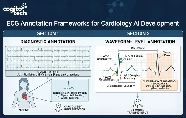
Medical waveform data annotation refers to the process of labeling these features with structured, time-aligned labels, including waveform segmentation, R-peak detection, interval marking, and cardiac event identification. Such annotation influences the fidelity of the learning and understanding of clinically relevant patterns within ECG signals by AI models, influencing their accuracy in clinical applications.
ECG Annotation: What Information Is Captured in an ECG Signal?
For AI models to interpret an ECG recording accurately, annotations must identify the individual components and events within the signal, rather than assigning only an overall diagnostic classification. ECG annotation captures the individual features and temporal relationships that define each cardiac cycle. This involves marking the onset and offset of P waves, QRS complexes, and T waves, identifying key fiducial points such as R-peaks, and recording measurements such as R-R intervals. Depending on the application, annotations may also identify individual beats, rhythm episodes, abnormal events, and sections affected by signal noise.
This creates an important distinction between diagnostic and waveform-level annotation. Diagnostic labels identify conditions such as atrial fibrillation or myocardial infarction, while waveform annotation pinpoints the exact locations and boundaries of the signal features associated with cardiac activity. Both provide different levels of information for cardiology AI development.
Why Accurate Medical Waveform Annotation Matters in Cardiology AI?
The utility of ECG annotation is not limited to describing ECG features and taking measurements. The accuracy and consistency of these annotations determine the quality of reference data used to develop and evaluate artificial intelligence models in cardiology. Medical waveform annotations directly affect the model learned during training, the accuracy of features extracted from ECG signals, and the reliability of performance estimates.
1. Model Training
Precisely marked waveform components give the AI model a consistent reference point for learning heart patterns. For instance, accurate identification of the boundaries of the P-wave and the QRS complex facilitates modeling of morphological variability, and correct identification of the R-peak supports learning of beat-to-beat timing patterns. Inconsistent annotations can introduce errors into these referenced models and affect what the model learns.
2. Feature Extraction
Several clinically relevant ECG measurements rely on the accurate recognition of the boundaries and peaks of the ECG waveforms. Annotations show high precision for the
PR interval: Measured from the onset of the P wave to the onset of the QRS complex.
QRS duration: The time interval between the beginning and end of the QRS complex.
QT interval: Measured from the onset of the QRS complex to the end of the T wave.
RR interval: Measured between two consecutive R-peaks.
These reference points, if incorrectly marked, will give an inaccurate measurement, which will impact the features used in subsequent analysis.
3. Model Evaluation
The accuracy of the annotations is also central to model evaluation of the reference data. A model can accurately recognize an abnormal rhythm but still give an incorrect boundary or interval measurement. To assess model performance, both diagnostic classification and waveform delineation, if applicable, should be evaluated.
Research Case Study: Improving ECG Waveform Delineation With Structured Segmentation
The need to evaluate waveform boundaries accurately is reflected in research on automated ECG delineation. For instance, the GLF-Net study investigates how cardiac cycle information can help AI models more accurately depict individual components of the ECG. The results are helpful for understanding the significance of structured segmentation in creating reliable ECG reference data.
Challenge
To accurately detect cardiac patterns, an AI model must also find start and end points of every component of a cardiac waveform. These boundaries are, however, not easy to recognize consistently because of the variations in the ECG morphology and differences between recordings. If reference boundaries are unknown or inconsistent, it becomes more difficult to train AI models and reliably evaluate their waveform predictions.
Approach
To solve these issues, the researchers created GLF-Net, a deep learning framework for ECG waveform segmentation. The approach is based on identifying R-peaks as reference points to segment the ECG signal into individual heartbeats. It then assigns signal points to four categories: P wave, QRS complex, T wave, and background. The model uses information about surrounding waveform patterns to help identify the boundaries of the waveforms based on their timing relationships with the cardiac components. The researchers tested its performance with two well-known ECG datasets, LUDB and QTDB.
Results
Evaluation results show that GLF-Net achieved an average F1 score of 97.60% on the LUDB dataset and 94.59% on the QTDB dataset. The F1 score measures how well a model’s predicted signal labels match the reference annotations, accounting for both false negatives and inaccurate predictions. These results demonstrate that the model was able to identify components of the ECG waveform with a high level of agreement with reference data in both datasets.
This result highlights why accurate waveform boundaries and consistently labeled reference data are important for the development of artificial intelligence in cardiology. If a model can be evaluated against reliable annotations, the ability of the model to identify individual cardiac components can be more meaningfully evaluated. Therefore, structured medical waveform annotations become an important part of creating and evaluating reliable datasets.
How ECG Annotation Services Support Cardiology AI Model Training and Evaluation
The study highlights the importance of consistent waveform boundaries and reference labels for building robust ECG AI models. The task of converting raw ECG recordings into AI training data requires workflows that capture the necessary signal features that models need to learn and ensure that the records are consistently labeled for validation and performance evaluation. The following are key annotation activities:
PQRST waveform delineation: Identification of the limits of P,Q,R,S and T wave components on the ECG.
Peak and fiducial-point annotation: Identifying R-peaks and other clinically relevant points for waveform analysis and interval measurements.
Wave onset/offset marking: Precisely recording where individual waveform components begin and end, particularly where boundaries are less distinct.
Rhythm labeling: Marking rhythm patterns, cardiac events, and other clinically relevant segments within the recording.
Multi-lead annotation: Maintain consistent annotations across multiple ECG leads while preserving temporal relationships between corresponding waveform features.
Quality review and adjudication: Review uncertain labels against defined guidelines and resolve disagreements through systematic adjudication.
Dataset formatting and organization: Structuring the completed annotations in consistent formats so they can be integrated into training, validation, and evaluation workflows.
Because waveform boundaries can vary in clarity across recordings, these activities need to follow well-defined labeling guidelines and review procedures. This helps maintain consistency throughout the ECG annotation services workflow and produces reference data that can be used more reliably across cardiology AI development.
What to Look for in Medical Waveform Annotation Companies
Choosing the correct partner for annotation entails assessing their capacity to correctly interpret complex physiological signals and ensure consistency in large datasets. The following are some important features to look for:
ECG-specific annotation expertise: Experience working with ECG recordings and understanding the requirements of cardiac waveform analysis.
PQRST waveform understanding: The ability to identify individual waveform components, their boundaries, peaks, and temporal relationships.
Peak and event identification: Precise recognition of R-peaks, R-R intervals, rhythm disturbances, and other clinically significant events.
Clear annotation guidelines: Defined guidelines for segmentation, event identification, and signal-quality assessment to minimize inconsistencies.
Quality assurance and expert validation: Review procedures with domain expertise for accuracy of annotations and interpretation of ambiguous waveform patterns.
Scalability: Processing large amounts of medical waveform data without losing consistency of annotations.
Complex pattern recognition: Experience identifying abnormal rhythms, variations in waveform morphology, and other clinically significant patterns.
Data security: Appropriate safeguards for protecting sensitive clinical information throughout the annotation process.
Cogito Tech’s medical waveform annotation services answer some of these questions. Our in- house team combines healthcare specialists and clinicians with data annotation experts to identify and classify physiological signal features. In ECG projects, we can provide P-QRS-T wave segmentation, R-R interval detection, peak detection, event marking, condition classification and signal quality labeling. Automated tools help detect and label baselines, while human-in-the-loop workflows support final clinical accuracy and context-relevant validation. The company also highlights data security and quality assurance for large volumes of data, which helps to build reference data for cardiology AI solutions.
The Road Ahead for Cardiology AI
Developing effective artificial intelligence in cardiology requires much more than large volumes of ECG recordings. Annotational quality, consistency and clinical relevance are similarly crucial to assist models in interpreting cardiac signals and providing relevant outputs. Each step ensures the data inputs for AI development are reliable, from defining the waves to structuring the data.Cogito Tech provides medical waveform annotation services covering ECG segmentation, peak detection, event labeling, and quality validation. Work with Cogito Tech to build precisely annotated, well-structured ECG datasets tailored to the requirements of your cardiology AI projects.
Frequently Asked Questions (FAQs)
What ECG features are typically captured through waveform annotation?+
ECG waveform annotation captures features such as P, QRS, and T wave boundaries, R-peaks, R-R intervals, individual beats, rhythm episodes, cardiac events, and signal-quality segments. Depending on the application, annotations can also include other fiducial points and clinically relevant waveform events.
Why is ECG annotation important for AI training?+
ECG annotation offers AI models with additional structured reference data, which helps them learn the pattern, morphology, and timing of cardiac waveforms. Accurate models that are well annotated enhance reliable training, validation, and evaluation of cardiology AI systems.
What does an ECG waveform represent?+
An ECG waveform is a graph that shows the electrical activity of the heart over time. The components of its waves (P, QRS, T) contain information about cardiac activity based on the duration, morphology and temporal relationship.
Why is precise ECG waveform annotation so important?+
Precise annotation ensures that waveform boundaries, peaks, intervals, and cardiac events are consistently identified. This affects the quality of the reference data used for AI model training and evaluation, supports accurate feature extraction, and helps determine how reliably a model identifies clinically relevant ECG patterns.
The post The Role of ECG Waveform Annotation in Training Cardiology AI appeared first on Cogitotech.